New preprint: Breaking new ground in brain imaging with three-photon microscopy
Our new preprint on Three-photon in vivo imaging of neurons and glia in the medial prefrontal cortex with sub-cellular resolutionꜛ is out! In our study, we showcase the power of three-photon microscopy to probe deeper into the brain than ever before, achieving remarkable imaging depth and resolution in live, behaving animals.
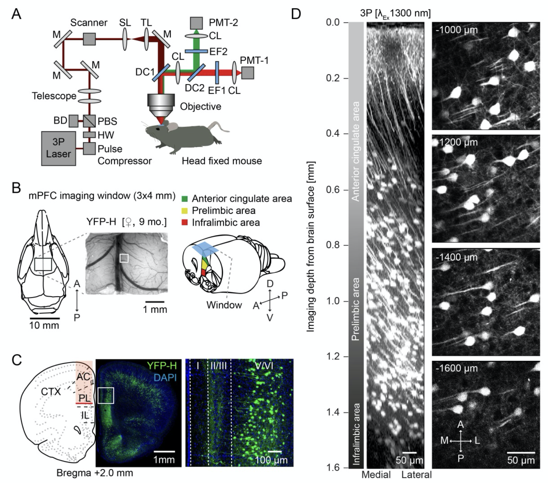 Fig. 1. from our preprintꜛ, showing the validation of 3P in vivo imaging of mPFC in a mouse model.
Update (May 2025): This preprint has now been peer-reviewed and published in Communications Biology:
Fuhrmann, Nebeling, Musacchio et al. (2025), Three-photon in vivo imaging of neurons and glia in the medial prefrontal cortex with sub-cellular resolution, https://www.nature.com/articles/s42003-025-08079-8ꜛ
Focus of our study
In our research, we targeted the medial prefrontal cortex (mPFC) in mice, a critical brain region involved in complex cognitive functions such as decision-making, memory, and emotional regulation. Additionally, we also investigated the hippocampus (hippocampal CA1 neurons) and spinal cord, regions that are typically challenging to access with conventional imaging techniques. Furthermore, we extended our imaging studies to Drosophila, utilizing three-photon microscopy to explore neural structures and activity in this model organism. This cross-species approach highlights the versatility of three-photon imaging and its applicability to a wide range of experimental models.
Animal models, brain regions, and methodologies
In mice, we focused on imaging the mPFC, including the prelimbic and infralimbic areas, as well as the hippocampus and spinal cord. These regions, located deep within the brain, are typically over 1 mm below the surface and have been largely inaccessible with previous imaging techniques. In Drosophila, we combined three-photon microscopy with non-invasive mounting methods to perform calcium imaging in the mushroom body Kenyon cells (KCs) through the intact cuticle, confirming the capability of three-photon imaging in small and complex organisms.
In mice, our three-photon setup enabled us to image structures as deep as 1.6 mm in the mPFC with sub-cellular resolution. This is a significant improvement over two-photon imaging, which reached only 800 µm under similar conditions. We could visualize neuronal activity and detailed structures such as dendritic spines and microglial processes at these depths.
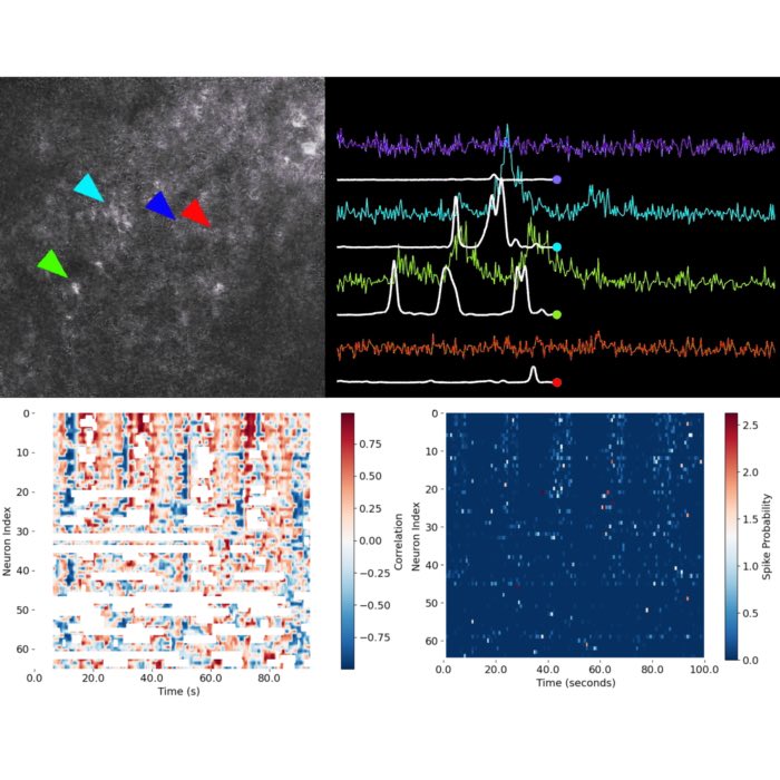
We recorded calcium transients from neurons and astrocytes up to 1.4 mm deep in the mPFC in awake, head-fixed mice. This allowed us to observe real-time brain activity during behavior. In Drosophila, we recorded neural activity at cellular resolution in the mushroom bodies, providing insights into the functioning of neural circuits in a different species.
In mice, we performed longitudinal imaging of dendritic spines on basal dendrites in the mPFC over a week, revealing their structural plasticity. We also imaged microglial processes at depths greater than 1 mm, quantifying their motility and observing stable turnover rates, even at these previously unreachable depths.
We also explored astrocytic calcium signaling in the mPFC, observing spontaneous calcium events in deep cortical layers (1.0-1.2 mm). These findings suggest a uniform astrocytic activity pattern across different cortical layers under anesthetized conditions.
Collaborative effort
This project was a highly collaborative effort, bringing together expertise from multiple research groups across different research institutes. The collaboration between the Neuroimmunology and Imaging Groupꜛ, the Axon Growth and Regeneration Groupꜛ, the Dynamics of Neuronal Circuits Groupꜛ, the Vascular Neurology Groupꜛ, and others was essential in advancing our understanding of three-photon imaging and its applications across various species and brain regions.
This work would also not have been possible without the support of the Core Research Facilities and Servicesꜛ and the Light Microscope Facilityꜛ at our research institute, the German Center for Neurodegenerative Diseases (DZNE)ꜛ.
Implications and future directions
Our findings demonstrate that three-photon microscopy is a powerful tool for investigating deep brain regions, such as the mPFC, without the need for invasive procedures like GRIN lens or microprism implantation. The successful application of this technique in both mice and Drosophila underscores its broad utility for neuroscience research.
The ability to image deep brain regions while animals are awake and behaving opens new avenues for exploring the neural circuits underlying cognition and behavior. This advancement will significantly enhance our understanding of brain function in both health and disease.
For a more detailed exploration of our methods and findings, feel free to read the full preprint hereꜛ.
Cite as
Please cite the preprint as follows:
Falko Fuhrmann, Felix C Nebeling, Fabrizio Musacchio, Manuel Mittag, Stefanie Poll, Monika Mueller, Eleonora Ambrad Giovannetti, Michael Maibach, Barbara Schaffran, Emily Burnside, Ivy Chi Wai Chan, Alex Lagurin, Nicole Reichenbach, Sanjeev Kaushalya, Hans-Ulrich Fried, Stefan Linden, Gabor Petzold, Gaia Tavosanis, Frank Bradke, Martin Fuhrmann, Three-photon in vivo imaging of neurons and glia in the medial prefrontal cortex with sub-cellular resolution, Commun Biol 8, 795 (2025), doi: 10.1038/s42003-025-08079-8ꜛ


comments